Description
Product Description
- The imaging principle of a fluorescence microscope is based on the physical phenomenon that certain molecules can absorb light of specific wavelengths (excitation light) and then emit light of other wavelengths (emission light). This technology is widely used in biological research to study the absorption, transport, distribution and localization of substances in cells, etc.
Fluorescence microscopes are mainly used to study samples such as organic and inorganic substances. Generally, fluorescence and phosphorescence are used to examine the structure, tissue and spatial distribution of samples. They are more suitable for studying complex samples that cannot be examined under traditional transmitted light microscopes.
Two different types of fluorescence microscopes:
Transmitted fluorescence microscope: Similar to ordinary optical microscopes, the light source starts from the base, passes through the condenser to the sample to excite fluorescence, then passes through the objective lens, and finally reaches the eyepiece.
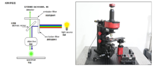
The other type is the most common epi-fluorescence microscope, which is adopted by most fluorescence microscopes. Its light source falls on the sample through the objective lens, excites fluorescence, and then emits back to the objective lens, finally reaching the eyepiece.
Technical Description
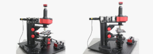
☑ What is a Fluorescence Microscope?
—— Experiment Summary:
Fluorescence microscopes are mainly used to study samples such as organic and inorganic substances. Generally, fluorescence and phosphorescence are used to examine the structure, tissue and spatial distribution of samples. They are more suitable for studying complex samples that cannot be examined under traditional transmitted light microscopes.
Two different types of fluorescence microscopes:
Transmitted fluorescence microscope: Similar to ordinary optical microscopes, the light source starts from the base, passes through the condenser to the sample to excite fluorescence, then passes through the objective lens, and finally reaches the eyepiece.
The other type is the most common epi-fluorescence microscope, which is adopted by most fluorescence microscopes. Its light source falls on the sample through the objective lens, excites fluorescence, and then emits back to the objective lens, finally reaching the eyepiece.
—— Working Steps:
The specific steps are as follows:
(1) Excitation Light Irradiation: Fluorescence microscopes use high-intensity light sources (such as xenon lamps, mercury lamps or lasers) to generate excitation light of specific wavelengths, which are guided to irradiate the sample through optical filters.
(2) Fluorescence Emission of the Sample: Fluorescent molecules (fluorescent dyes or fluorescent proteins) in the sample absorb the energy of the excitation light and enter an excited state. Subsequently, they return to the ground state by emitting light of a longer wavelength, and the light generated in this process is called fluorescence.
(3) Filtering and Imaging: The emitted fluorescence passes through the emission filter of the microscope, which filters out the remaining excitation light and only allows fluorescence of specific wavelengths to pass through. Then, the detection system of the microscope (such as an eyepiece or a camera) captures these emitted lights to generate a clear sample image.
—— Experimental Objectives:
Based on the fluorescence phenomenon, that is, certain substances emit light of different wavelengths after absorbing light of specific wavelengths. This technology is widely used in biological research to study the absorption and transport of substances in cells, the distribution and localization of chemical substances, etc.
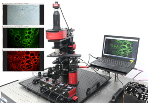
(1) Specific Molecular Labeling and Observation: Through fluorescence labeling technology, specific molecules or structures in the sample, such as proteins, DNA or RNA, can be observed. With the help of fluorescent probes, researchers can directly observe the distribution and dynamic changes of these molecules.
(2) High-Contrast Imaging: Fluorescence microscopes have an extremely high signal-to-noise ratio. Unlabeled parts in the sample usually do not emit light, which reduces background interference and significantly improves the visibility and contrast of target molecules or structures.
(3) Live Cell Dynamic Imaging: Fluorescence microscopes can be used to observe real-time dynamic processes of living cells or tissues, such as cell division, substance transport, signal transduction, etc.
(4) Multiple Labeling and Multicolor Imaging: Using multiple fluorescent dyes of different wavelengths, fluorescence microscopes can simultaneously label and observe multiple different biological targets, revealing their relative positions and interactions in the sample.
(5) Three-Dimensional Imaging: Fluorescence microscopes can be combined with other technologies (such as confocal microscopes) to construct three-dimensional images of samples by scanning fluorescent signals from different focal planes.
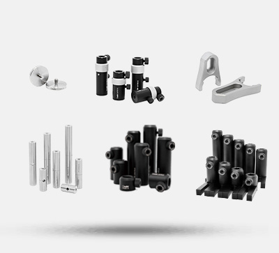
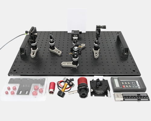
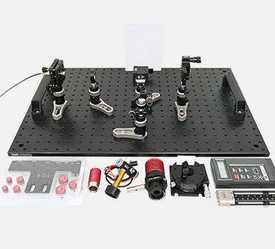
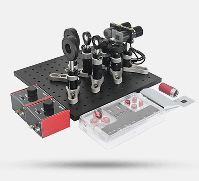
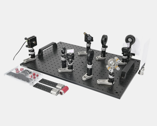
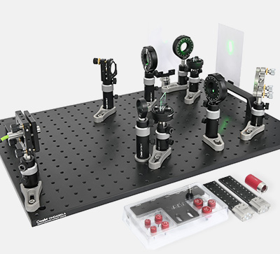
—— Kit List:
For detailed information on the detailed list of this kit, please contact customer service.

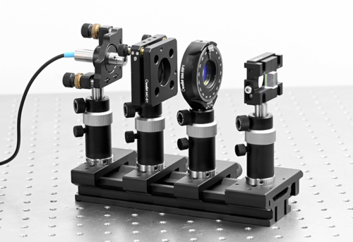
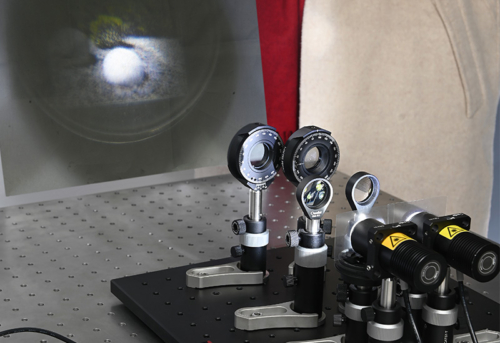
—— Experimental Content
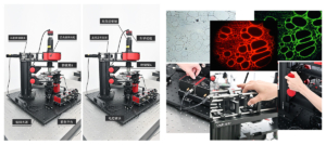
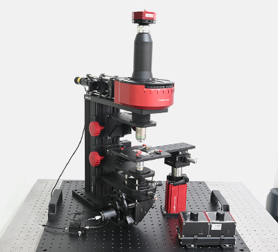
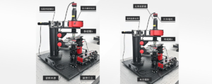
Assembly
Application Examples